
Quiz Summary
0 of 86 questions completed
Questions:
Information
You have already completed the quiz before. Hence you can not start it again.
Quiz is loading…
You must sign in or sign up to start the quiz.
You must first complete the following:
Results
Results
0 of 86 questions answered correctly
Your time:
Time has elapsed
You have reached 0 of 0 point(s), (0)
Earned Point(s): 0 of 0, (0)
0 Essay(s) Pending (Possible Point(s): 0)
Categories
- FillInTheBlank 0%
- Multiple Choice 0%
- Summary 0%
- 1
- 2
- 3
- 4
- 5
- 6
- 7
- 8
- 9
- 10
- 11
- 12
- 13
- 14
- 15
- 16
- 17
- 18
- 19
- 20
- 21
- 22
- 23
- 24
- 25
- 26
- 27
- 28
- 29
- 30
- 31
- 32
- 33
- 34
- 35
- 36
- 37
- 38
- 39
- 40
- 41
- 42
- 43
- 44
- 45
- 46
- 47
- 48
- 49
- 50
- 51
- 52
- 53
- 54
- 55
- 56
- 57
- 58
- 59
- 60
- 61
- 62
- 63
- 64
- 65
- 66
- 67
- 68
- 69
- 70
- 71
- 72
- 73
- 74
- 75
- 76
- 77
- 78
- 79
- 80
- 81
- 82
- 83
- 84
- 85
- 86
- Current
- Review
- Answered
- Correct
- Incorrect
- Question 1 of 86
1. Question
Transmission of sound
What is transmission of sound in a medium due to? CorrectIncorrect
 - Question 2 of 86
2. Question
Sound wave characteristics
What is Hertz the SI unit of?CorrectIncorrect - Question 3 of 86
3. Question
Attenuation
Insert the correct word(s) to complete the statement: Attenuation depends on the ….. of the sound wave.(There may be more than one correct answer)CorrectIncorrect - Question 4 of 86
4. Question
Transducer orientation
The image shows correct and inverted orientation of the probe. Which of the following statements are correct? Select all that apply.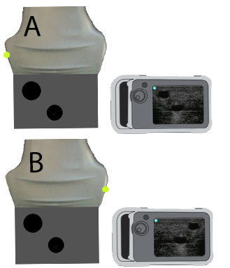 CorrectIncorrectQuestion 5 of 86
5. Question
Image optimisation A
The image shows two ultrasound images, where B is an optimized image of A. How is B optimized ?CorrectIncorrectQuestion 6 of 866. Question
Image optimisation B
The image shows two ultrasound images, where B is an optimized image of A. How is B optimized ?CorrectIncorrectQuestion 7 of 867. Question
Position 1 – orientation
The image on the left shows two presentations on the screen of the subcostal 4-chamber view. Which of the images, A or B, is oriented correctly?CorrectIncorrectQuestion 8 of 868. Question
FATE views
The image on the left shows the four cardiac FATE views. Which of the following statements is correct?CorrectIncorrectQuestion 9 of 869. Question
Key points regarding the subcostal 4 chamber view
Which statements regarding evaluating the heart by the subcostal 4 chamber view are true? Select all that apply.CorrectIncorrectQuestion 10 of 8610. Question
Key points of apical 4 chamber view
Which of the following statements about the apical 4 chamber view is correct?CorrectIncorrectQuestion 11 of 8611. Question
Key points of the PLAX view
What is true about evaluating the heart by the PLAX view? Select all that apply.CorrectIncorrectQuestion 12 of 8612. Question
Image presentation
Two conventions exist regarding the orientation indicator on the screen. Which of these statements are correct concerning the FATE examination (cardiac ultrasound)?CorrectIncorrectQuestion 13 of 8613. Question
Pathology 1A. The video clips show four PSAX views. Which one represents significant pathology (only one “True”) ?
CorrectIncorrectQuestion 14 of 8614. Question
Pathology 1B. The video clip shows one PSAX view. Which one of the statements below is correct (only one “True”) ?
CorrectIncorrectQuestion 15 of 8615. Question
Pathology 2A. The video clips show four SC4CH views. Which one represents significant pathology (only one “True”) ?
CorrectIncorrectQuestion 16 of 8616. Question
Pathology 3A. The video clips show four PLAX views. Which one represents significant pathology (only one “True”) ?
CorrectIncorrectQuestion 17 of 8617. Question
Pathology 3B. The video clip shows one PLAX view. Which one of the statements below is correct (only one “True”) ?
CorrectIncorrectQuestion 18 of 8618. Question
Pathology 4A. The video clips show four AP4CH views. Which one represents significant pathology (only one “True”) ?
CorrectIncorrectQuestion 19 of 8619. Question
Pathology 4B. The video clip shows one AP4CH view. Which one of the statements below is correct (only one “True”) ?
CorrectIncorrectQuestion 20 of 8620. Question
Lung-pleura. Which of these statements are correct? Select all that apply.CorrectIncorrectQuestion 21 of 8621. Question
Pathology 5A. The video clips show four PLAX views. Which one represents significant pathology (only one “True”) ?
CorrectIncorrectQuestion 22 of 8622. Question
Pathology 5B. The video clip shows one PLAX view. Which one of the statements below is correct (only one “True”) ?
CorrectIncorrectQuestion 23 of 8623. Question
Pathology 6A. The video clips show four AP4CH views. Which one represents significant pathology (only one “True”) ?
CorrectIncorrectQuestion 24 of 8624. Question
Pathology 6B. The video clip shows one AP4CH view. Which one of the statements below is correct (only one “True”) ?
CorrectIncorrectQuestion 25 of 8625. Question
Pathology 7A. The video clips show four S4CH views. Which one represents significant pathology (only one “True”) ?
CorrectIncorrectQuestion 26 of 8626. Question
Pathology 7B. The video clip shows one S4CH view. Which one of the statements below is correct (only one “True”)?
CorrectIncorrectQuestion 27 of 8627. Question
Pathology 8A. The video clips show four PLAX views. Which one represents significant pathology (only one “True”)?
CorrectIncorrectQuestion 28 of 8628. Question
Pathology 8B. The video clip shows one PLAX view. Which one of the statements below is correct (only one “True”)?
CorrectIncorrectQuestion 29 of 8629. Question
Pathology 9A. The video clip shows four SC4CH views. Which one represents significant pathology (only one “True”)?
CorrectIncorrectQuestion 30 of 8630. Question
Pathology 9B. The video clip shows one SC4CH view. Which one of the statements below is correct (only one “True”)?
CorrectIncorrectQuestion 31 of 8631. Question
Pathology 10A. The video clip shows four AP4CH views. Which one represents significant pathology (only one “True”)?
CorrectIncorrectQuestion 32 of 8632. Question
Pathology 10B. The video clip shows one AP4CH view. Which one of the statements below is correct (only one “True”)?
CorrectIncorrectQuestion 33 of 8633. Question
Pathology 11A. The video clips show four PSAX views. Which one represents significant pathology (only one “True”)?
CorrectIncorrectQuestion 34 of 8634. Question
Pathology 11B. The video clip shows one PSAX view. Which one of the statements below is correct (only one “True”)?
CorrectIncorrectQuestion 35 of 8635. Question
Pathology 12A. The video clip shows four IVC views. Which one of the statements below is correct (only one “True”)?
CorrectIncorrectQuestion 36 of 8636. Question
Pathology 12B. The video clip shows one subcostal view. Which one of the statements below is correct (only one “True”)?
CorrectIncorrectQuestion 37 of 8637. Question
Pathology 13A. The video clip shows four S4CH views. Which one represents significant pathology (only one “True”)?
CorrectIncorrectQuestion 38 of 8638. Question
Pathology 13B. The video clip shows one S4CH view. Which one of the statements below is correct (only one “True”)?
CorrectIncorrectQuestion 39 of 8639. Question
Pathology 14A. The video clips show four PLAX views. Which one represents significant pathology (only one “True”)?
CorrectIncorrectQuestion 40 of 8640. Question
Pathology 14B. The video clip shows one PLAX view. Which one of the statements below is correct (only one “True”)?
CorrectIncorrectQuestion 41 of 8641. Question
Pathology 15A. The video clips show four A4CH views. Which one represents significant pathology (only one “True”)?
CorrectIncorrectQuestion 42 of 8642. Question
Pathology 15B. The video clip shows one A4CH view. Which one of the statements below is correct (only one “True”)?
CorrectIncorrectQuestion 43 of 8643. Question
Pathology 16A. The video clips show four PSAX views. Which one represents significant pathology (only one “True”)?
CorrectIncorrectQuestion 44 of 8644. Question
Pathology 16B. The video clip shows one PSAX view. Which one of the statements below is correct (only one “True”)?
CorrectIncorrectQuestion 45 of 8645. Question
Pathology 17A. The video clip shows four A4CH views. Which one represents significant pathology (only one “True”)?
CorrectIncorrectQuestion 46 of 8646. Question
Pathology 17B. The video clip shows one A4CH view. Which one of the statements below is correct (only one “True”)?
CorrectIncorrectQuestion 47 of 8647. Question
Pathology 18A. The video clips show four S4CH views. Which one represents significant pathology (only one “True”)?
CorrectIncorrectQuestion 48 of 8648. Question
Pathology 18B. The video clip shows one S4CH view. Which one of the statements below is correct (only one “True”)?
CorrectIncorrectQuestion 49 of 8649. Question
Pathology 19A. The video clips show four PSAX views. Which one represents significant pathology (only one “True”)?
CorrectIncorrectQuestion 50 of 8650. Question
Pathology 19B. The video clip shows one PSAX view. Which one of the statements below is correct (only one “True”)?
CorrectIncorrectQuestion 51 of 8651. Question
Pathology 20A. The video clip shows four pleural views. Which one of the statements below is correct (only one “True”)?
CorrectIncorrectQuestion 52 of 8652. Question
Pathology 20B. The video clip shows one pleural view. Which one of the statements below is correct (only one “True”)?CorrectIncorrectQuestion 53 of 8653. Question
Pathology 21A. The video clip shows four IVC views. Which one of the statements below is correct (only one “True”)?
CorrectIncorrectQuestion 54 of 8654. Question
Pathology 21B. The video clip shows one IVC view. Which one of the statements below is correct (only one “True”)?
CorrectIncorrectQuestion 55 of 8655. Question
Pathology 22A. The video clip shows four 4CH views. Which one of the statements below is correct (only one “True”)?
CorrectIncorrectQuestion 56 of 8656. Question
Pathology 22B. The video clip shows one 4CH view. Which one of the statements below is correct (only one “True”)?
CorrectIncorrectQuestion 57 of 8657. Question
Pathology 23A. The video clip shows four pleural views. Which one of the statements below is correct (only one “True”)?
CorrectIncorrectQuestion 58 of 8658. Question
Pathology 23B. The video clip shows one pleural view. Which one of the statements below is correct (only one “True”)?
CorrectIncorrectQuestion 59 of 8659. Question
Pathology 24A. The video clip shows four pleural views. Which one of the statements below is correct (only one “True”)?
CorrectIncorrectQuestion 60 of 8660. Question
Pathology 24B. The video clip shows one pleural view. Which one of the statements below is correct (only one “True”)?
CorrectIncorrectQuestion 61 of 8661. Question
Pathology 25A. The video clip shows four PLAX views. Which one of the statements below is correct (only one “True”)?
CorrectIncorrectQuestion 62 of 8662. Question
Pathology 25B. The video clip shows one PLAX view. Which one of the statements below is correct (only one “True”)?
CorrectIncorrectQuestion 63 of 8663. Question
Recognising veins and arteries on an ultrasound image. Look at the three video recordings of veins and arteries. Which of the following statements are correct? Select all that apply.
CorrectIncorrectQuestion 64 of 8664. Question
Diameter of veins. The image displays several veins. Which of the following statements are correct? Select all that apply.
CorrectIncorrectQuestion 65 of 8665. Question
Appropriate length of an intravenous catheter. Which of the following statements regarding length of intravascular catheters are true? Select all that apply.
CorrectIncorrectQuestion 66 of 8666. Question
Needle guidance
Which statements regarding needle guidance are correct? Select all that apply.CorrectIncorrectQuestion 67 of 8667. Question
Needle guidance and skin puncture
Which statements are regarding needle guidance and skin puncture are correct? Select all that apply.CorrectIncorrectQuestion 68 of 8668. Question
Important points for successful cannulation. Which statements about cannulation are correct? Select all that apply.CorrectIncorrectQuestion 69 of 8669. Question
2D sonoanatomy of the cardiac structures – position 1
In the image each letter labels a structure. Insert the correct letters for each statement and click Next.- shows RA = right atriumshows the livershows the apexshows RV = right ventricleshows LV = left ventricleshows LA = left atrium
Correct 30 / 30 PointsIncorrect / 30 PointsQuestion 70 of 8670. Question
Test 1: 2D sonoanatomy of the cardiac structures
Test yourself: Insert missing letters. In the image each letter labels a structure. Please compare the image with the statements. Insert the correct letter missing in each statement and click the ‘Next- Shows LA = Left atriumShows Descending thoracic aortaShows RA = Right atriumShows RV = Right ventricleShows LV = Left ventricleShows Apex
Correct 5 / 5 PointsIncorrect / 5 PointsQuestion 71 of 8671. Question
2D sonoanatomy of the cardiac structures
In the image each letter labels a structure. Insert the correct letters for each statement and click Next.- shows IAS: inter-atrial septumshows the anterior mitral leafletshows IVS: inter-ventricular septumshows the lateral wallshows the posterior mitral leafletshows the base of the heart
Correct 30 / 30 PointsIncorrect / 30 PointsQuestion 72 of 8672. Question
2D sonoanatomy of the cardiac structures – PLAX
In the image each letter labels a structure. Insert the correct letters for each statement and click Next.- shows the posterior wallshows the descending aortashows the anterior mitral leafletshows the left atriumshows the pericardiumshows the right ventricle
Correct 30 / 30 PointsIncorrect / 30 PointsQuestion 73 of 8673. Question
2D sonoanatomy of the cardiac structures – PSAX
In the image each letter labels a structure. Insert the correct letters for each statement and click Next.- shows the right ventricleshows the left ventricleshows the pericardiumshows the posterior wallshows the lateral wallshows the papillary muscle
Correct 30 / 30 PointsIncorrect / 30 PointsQuestion 74 of 8674. Question
Sonoanatomy of the right pleura
In the image each letter labels a structure. Insert the correct letters for each statement and click Next.- shows the livershows the caudal directionshows the orientation indicatorshows the spleenshows the cranial directionshows the diaphragm
Correct 30 / 30 PointsIncorrect / 30 PointsQuestion 75 of 8675. Question
2D sonoanatomy of the RUQ and LUQ views
In the image each letter labels a structure. Insert the correct letters for each statement and click Next.- shows the spleenshows Morison’s pouchshows the livershows the diaphragm on patient’s right sideshows the diaphragm on patient’s left sideshows splenorenal recess
CorrectIncorrectQuestion 76 of 8676. Question
Identifying the structures on the ultrasound image of the chest wall
In the image each letter labels a structure. Insert the correct letters for each statement and click “Next”.- shows skinshows image artefacts from the echoes of the overlying skin, muscle, costae and pleural lineshows rib shadowshows costashows intercostal muscleshows pleura
CorrectIncorrectQuestion 77 of 8677. Question
Identifying the structures
In the image each letter labels a structure. Insert the correct letters for each statement and click “Next”.- shows the spleenshows the diaphragm on the left sideshows the diaphragm on the right sideshows the area corresponding to left lungshows the area corresponding to right lungshows the liver
CorrectIncorrectQuestion 78 of 8678. Question
Identifying structures in pleural effusion
In the image each letter labels a structure. Insert the correct letters for each statement and click “Next”.- shows the spleenshows the pleural effusion between right lung and right diaphragmshows the pleural effusion between left lung and left diaphragmshows the diaphragm on the right sideshows the diaphragm on the left sideshows the liver
CorrectIncorrectQuestion 79 of 8679. Question
Equipment and skills summary
Match the phrase at the top with the correct option below to form true statements by dragging the answer into the answer box.Sort elements
- is abbreviated OM
- gives excellent high resolution 2D-images in superficial structure
- is on the right side of the screen
- is on the left side of the screen
- adjust gain at different depths of the ultrasound image
- permit deep penetration in patients
- The orientation marker on the transducer
- High frequencies linear transducers
- In cardiac ultrasound the orientation indicator (OI)
- In FAST ultrasound the orientation indicator (OI)
- Time gain compensation (TGC)
- Curved array (abdominal) probes
CorrectIncorrectQuestion 80 of 8680. Question
Match the phrases on the left side with the phrases on the right side to form true statements. Click “Submit” when finishedhttps://test.usabcd.org/wp-content/uploads/FM_media/BasUS_studmed/BasUS_studmed_Q_M7_P15?
Sort elements
- Left atrium
- Right atrium
- Left ventricle
- Right ventricle
- Aorta
- LA is the abbreviation for
- RA is the abbreviation for
- LV is the abbreviation for
- RV is the abbreviation for
- AO is the abbreviation for
CorrectIncorrectQuestion 81 of 8681. Question
Match the phrase at the top with the correct option below to form true statements by dragging the answer into the answer box.
Sort elements
- The subcostal 4-chamber view, and is obtained from position 1 on the FATE card
- The apical 4-chamber view, and is obtained from position 2 on the FATE card
- The parasternal long-axis view, and is obtained from position 3 on the FATE card
- The parasternal short-axis view, and is obtained from position 3 on the FATE card
- Obtained from position 4 on the FATE card
- S4CH view is
- A4CH view is
- PLAX view is
- PSAX view is
- Pleura view is
CorrectIncorrectQuestion 82 of 8682. Question
Match the phrases on the left side with the phrases on the right side to form true statements. Click “Submit” when finishedhttps://test.usabcd.org/wp-content/uploads/FM_media/BasUS_studmed/BasUS_studmed_Q_M7_P43?
Sort elements
- the subcostal or subxiphoid view
- directed towards the patients LEFT.
- will appear in the near field of the image
- will appear in the far field of the image
- will appear on the right side of the screen
- The FATE projection 1 is called
- In the FATE projection 1 the orientation marker on the transducer should be
- On the ultrasound image the RA and RV
- On the ultrasound image the LA and LV
- In the subcostal 4 chamber view the apex of the heart
CorrectIncorrectQuestion 83 of 8683. Question
Match the phrases on the left side with the phrases on the right side to form true statements. Click “Submit” when finishedhttps://test.usabcd.org/wp-content/uploads/FM_media/BasUS_studmed/BasUS_studmed_Q_M7_P57?
Sort elements
- the apical 4 chamber view (A4CH)
- directed towards the patients left shoulder and backwards
- will appear in the near field of the image
- is seen behind the left atrium
- is a good starting point for the apical 4 chamber view examination
- The FATE position 2 is called
- In the FATE position 2 the orientation marker on the transducer should be
- Both right and left ventricle
- The descending thoracic aorta
- The ictus cordis
CorrectIncorrectQuestion 84 of 8684. Question
Match the phrases on the left side with the phrases on the right side to form true statements. Click “Submit” when finishedhttps://test.usabcd.org/wp-content/uploads/FM_media/BasUS_studmed/BasUS_studmed_Q_M7_P88?
Sort elements
- are fixed points and should always be identified
- appears above the liver
- is on the right side of the screen
- if the orientation indicator is on the right side of the screen
- both on the left and right side
- The right and left diaphragms
- The diaphragm on the right side
- In echocardiography the orientation indicator
- In pleural scan with the FATE protocol (position 4) the orientation marker is generally pointed cranially
- Pleura should be examined
CorrectIncorrectQuestion 85 of 8685. Question
How to scan – aorta ultrasound
Match the phrase at the top with the correct option below to form true statements by dragging the answer into the answer box.Sort elements
- a cross sectional view
- a longitudinal view
- the right side of the screen
- the left side of the screen
- 15 cm
- 5 MHz
- First scan from the epigastrium to the umbilicus with
- Then turn the probe clockwise and scan from the bifurcation to the celiac trunk with
- In the horizontal plane: Display the left side of the patient on
- In the longitudinal plane: Display the cranial end of the patient on
- Initial depth should be adjusted to
- Select a curved array probe scanning up to
CorrectIncorrectQuestion 86 of 8686. Question
Sonopathology and measurement
Match the phrase at the top with the correct option below to form true statements by dragging the answer into the answer box.Sort elements
- anterior-posterior direction
- left-right direction
- outer wall to outer wall
- inner wall to inner wall of the aneurysmal lumen
- perpendicular to the long axis of the vessel
- perpendicular to the skin surface
- Measure the aortic diameter in the
- Do not measure the aortic diameter in the
- Gauge the diameter of the aneurysm from
- Never gauge the diameter of the aneurysm from
- Always gauge the diameter of the aortic vessel
- The long axis of the abdominal aorta is not always
CorrectIncorrect